OCT

Optical computed tomography (OCT) or optical coherence tomography, is a non-invasive diagnostic exam which allows obtaining scansions of the cornea and retina for the diagnosis and follow-up of numerous corneal and retinoic diseases, and for the preoperative diagnosis and postoperative follow-up of most eye diseases needing surgery.
What is optical computed tomography (OCT)?
It is a non-invasive imaging technique, based on white light or low coherency interferometry, a Laser bundle with no harmful radiations, which is used to analyze ocular structures, especially retinoic and corneal, by means of high resolution sections.
What is optical computed tomography (OCT) used for?
OCT allows obtaining very precise corneal and retinoic scansions and subsequently a detailed analysis of corneal strata, the central region of the retina called the macula, and the optic nerve. This “imaging” method allows a doctor to diagnose and monitor numerous corneal and retinoic diseases, such as, for example, senile macular degeneration, diabetic retinopathy and glaucoma. It is particularly useful in cases of macular edema of various origin. OCT is an indispensable exam in the preoperatory diagnosis and postoperative follow-up of most eye diseases needing surgery. Since it is a digitalized exam, it allows comparing different tests thus providing differential maps of the patient’s eyes. It is a fundamental exam in the early diagnosis of some diseases: for example, in patients affected by glaucoma, OCT is able to measure the thickness of the nervous fibers surrounding the optic nerve, showing, in some cases, an early alteration of said fibers in the presence of a normal visual field, and this allows initiating promptly a treatment in order to slow down the progression of the disease.
Who can undergo optical computed tomography (OCT)?
All patients in whom a corneal, retinoic or optic nerve disease is suspected, except for those presenting with a remarkable opacity of the crystalline lens, important modifications of the lacrimal film and absence of eye fixation.
Is optical computed tomography (OCT) painful or dangerous?
It is a reliable, non-invasive, harmless exam, not involving any contact.
How does optical computed tomography (OCT) work?
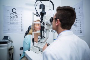
Performing an OCT is simple and fast, and lasts about 10-15 minutes. The patient is seated in front of the device and is invited by the operator to fix a luminous point with his/her eye: scansion begins immediately when the eye structure to be analyzed is focused. With last generation OCTs, the exam may be performed even without dilating the pupil, after the operator has evaluated the features of the eye and the type of disease to be investigated.
